出版社: Investigative Ophthalmology & Visual Science November 2025, Vol.66, 42. doi:https://doi.org/10.1167/iovs.66.14.42

作者: Fan Xia; Shuizhen Shi; Erick Palacios; Won-Kyu Ju; Hua Liu; Wenbo Zhang

CXCR3 缺失通过调控青光眼小鼠模型中的神经元‑星形胶质细胞通讯减轻视网膜神经节细胞丢失

研究背景:

青光眼是以进行性视网膜神经节细胞(RGC)丢失和视神经(ON)变性为主要特征的多因素神经退行性疾病,也是全球不可逆性失明的第二大病因。

青光眼进展与眼压(IOP)升高密切相关,眼压也是目前研究最为深入、且唯一可干预的青光眼危险因素。然而,仅降低眼压并不能完全阻止青光眼病情进展。

视网膜神经节细胞的丢失具有不可逆性。阐明其损伤机制并寻找可预防神经节细胞丢失的干预手段,仍是目前亟待解决的关键问题。利用微球诱导的青光眼模型,我们进一步证实:CXCL10/CXC 趋化因子受体 3(CXCR3)信号通路通过调控补体成分 3(C3)/C3a 受体(C3aR)通路介导的神经元‑星形胶质细胞相互作用,在视网膜神经节细胞(RGC)丢失中发挥关键作用。本研究对青光眼小鼠模型的公共视网膜转录组测序(RNA‑seq)数据集进行了重新分析,重点关注趋化因子表达的变化。

方法

我们重新分析了一份公开发表的青光眼小鼠模型视网膜整体转录组测序(RNA-seq)数据集,以筛选差异表达的趋化因子。采用微球诱导的小鼠青光眼模型、原代视网膜神经节细胞(RGC)及星形胶质细胞,探讨CXC 趋化因子配体 10(CXCL10)/CXC 趋化因子受体 3(CXCR3)轴在疾病中的作用。

对8~10 周龄小鼠采用微球注射法构建青光眼模型:小鼠经腹腔注射 75 mg/kg 苯巴比妥钠麻醉,予0.5% 盐酸丙美卡因表面麻醉,使用1% 托吡卡胺与 2.5% 盐酸去氧肾上腺素散瞳。用27G 针头制作角膜隧道,再以32G 针头及微量进样器(Hamilton Company, Reno, NV, USA)向前房内依次注入:

  • 2 µL 直径 1 µm 聚苯乙烯微球悬液(含 3.0×10⁷ 个微球;Polysciences, Warrington, PA, USA)
  • 2 µL 直径 6 µm 聚苯乙烯微球悬液(含 6.3×10⁶ 个微球;Polysciences)
  • 1 µL 含 30% Healon 的磷酸盐缓冲液(PBS)。

该方法可使眼压中度升高并至少维持 6 周。于每日15:00–17:00使用大小鼠专用回弹式眼压计Tonolab(Icare Finland Oy,芬兰赫尔辛基)在固定时间点测量眼压,以减小昼夜波动干扰。

自微球注射后第 3 天开始,每 12 小时腹腔注射一次 C3aR 拮抗剂 SB290157(20 mg/kg)或溶媒对照(含 10% Tween 80 和 10% 乙醇的 PBS),连续给药 6 周。

结果

在疾病进展过程中,包括CXCL10在内的多种趋化因子显著上调,该因子既往已被证实与神经变性相关。在微球诱导模型中,注射后第 5 天CXCL10表达即显著升高。

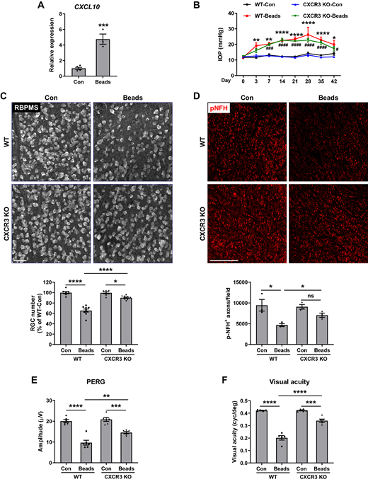
术后 6 周时,敲除 CXCL10 的受体CXCR3可在不影响眼压(IOP)的前提下,显著减轻视网膜神经节细胞(RGC)丢失与轴突变性。通过图形视网膜电图及视力检测发现,CXCR3 敲除小鼠的视觉功能得到保留。

机制上,CXCL10/CXCR3 信号通路可上调星形胶质细胞中补体成分 3(C3)及 RGC 中C3a 受体(C3aR)的表达,介导具有损伤作用的星形胶质细胞–RGC 交互对话。

通过玻璃体内注射腺相关病毒介导的显性负性 CXCL10进行基因治疗,或药物阻断 C3aR,均可有效减少 RGC 丢失。

图一
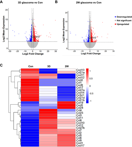
Transcriptomic profiling of GSE241782 identified upregulation of several chemokines, including CXCL10, in glaucomatous retina. (A, B) Volcano plots of upregulated and downregulated genes in glaucomatous versus control retinas at 3 days (3D) and 2 weeks (2W) following microbead injection. CXCL10 is prominently upregulated at both time points. Each experimental group had four replicate samples; sexes were pooled for n = 2 RNA-seq libraries. (C) Heatmap of differentially expressed chemokine transcripts across glaucoma progression (control, 3D, and 2W).

图二

 CXCR3 deletion was neuroprotective in the microbead-induced glaucomatous retina. (A) CXCL10 mRNA was measured at 5 days after microbead injection (n = 3–5). ***P < 0.001. (B) IOP of sham and microbead-injected eyes of WT and CXCR3 KO mice was measured at various time points (n = 10–12). *P < 0.05, **P < 0.01, ****P < 0.0001 compared with WT control (WT-Con); #P < 0.05, ###P < 0.001, and ####P < 0.0001 compared with CXCR3 KO control (KO-Con). (C) Retinal flatmounts were stained with anti-RBPMS to label RGCs at 6 weeks after microbead injection. Bar graph represents the quantification of  RGC number. RGC numbers from eight images of the peripheral retina were counted and averaged as one sample (n = 7–9). (D) Optic nerve sections at 6 weeks after microbead injection were stained with anti-pNFH (red) to visualize axons. Bar graph represents the quantification of pNFH+ axons. Axon numbers from three to five images were counted and averaged as one sample (n = 3). (E, F) PERG (n = 5, 6) and visual acuity (n = 5 or 6) were measured at 6 weeks after microbead injection. Scale bar: 50 µm. *P < 0.05, **P < 0.01, ***P < 0.001, ****P < 0.0001; ns, not significant.

结论

CXCL10/CXCR3 信号通路是青光眼视网膜神经节细胞(RGC)丢失的关键介导因子。靶向该通路及其下游上调的 C3/C3aR 信号轴,有望成为一种 ** 不依赖于眼压(IOP)** 的青光眼治疗新策略。